
Renal Angiomyolipoma
Renal angiomyolipoma embolization is a benign tumor in the kidney, occurring in 0.2-0.6% of the population.1 Structurally, angiomyolipomas consist of three components – blood vessels (‘angio’), smooth muscle (‘myo’), and fat (adipose tissue; ‘lipo’) – in varying proportions.1
Prevalence statistics suggest that renal angiomyolipomas are twice as likely to form in women than in men.1-3 Moreover, in 20% of the cases, renal angiomyolipomas coincide with the occurrence of the rare genetic disease tuberous sclerosis complex.2
Renal Angiomyolipoma: Symptoms
Renal Angiomyolipoma: Risks
While renal angiomyolipomas are benign, they are not without risks. Generally, renal angiomyolipomas pose a considerable risk of hemorrhage (bleeding) due to the possibility of tumor rupture.3,4 The occurrence of angiomyolipoma hemorrhage can be life-threatening.4 In rare cases, renal angiomyolipomas may also lead to chronic kidney disease.5
Renal angiomyolipomas in pregnant women pose an even higher risk.4 They grow more quickly, become invasive, and are more likely to rupture and cause hemorrhage.4 It is important to note, however, that angiomyolipomas with a low fat content are typically low-risk.3
Renal Angiomyolipoma: Diagnosis
Diagnosis of renal angiomyolipomas most commonly takes place through imaging.1 While it is straightforward to diagnose renal angiomyolipomas with a high fat content, diagnosis of low-fat angiomyolipomas can be challenging.2,3 Imaging modalities such as computed tomography (CT) and magnetic resonance imaging (MRI) easily confirm the presence of renal angiomyolipoma when fat is aggregated in bulk quantities.3
However, at least 5% of the cases of renal angiomyolipoma lack enough fat for CT or MRI alone to be able to confirm a diagnosis.3 Using both CT and MRI in combination is a good strategy for diagnosing low-fat angiomyolipomas.3 Alternatively, immunohistochemical methods can be used to establish the presence of low-fat renal angiomyolipomas.3
Renal Angiomyolipoma: Treatment Options
Surgical Options
Non-Surgical Option
Selective renal artery embolization of the angiomyolipoma is a preventive outpatient treatment approach that is now the standard treatment for all sizes of angiomyolipoma. This involves placing a small tube in an artery in the leg or arm and navigating a small catheter into a branch of the renal artery near the tumor site.5,6 Small blocking agents, such as polyvinyl alcohol particles, are then injected into the blood vessels to cut off tumor blood supply.5,6 The tumor then becomes inactive thus disabling any chance of tumor rupture or bleeding.
Angiomyolipoma Non-Surgical Embolization: What to Expect
Selective embolization is an outpatient non-surgical procedure with minimal downtime. The procedure is done in an outpatient state of the art center where our interventional radiologist performs the treatment through a tiny tube called a catheter. This procedure can be performed by either placing the catheter in an artery at the top of the leg (called a femoral approach) or by placing it into an artery in the lower arm (called a radial approach). The two methods are illustrated below.
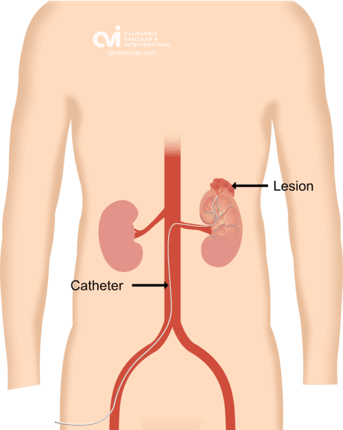
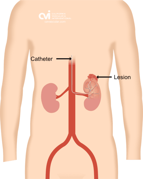
The patient is given a local and topical anesthetic to numb the skin and a mild sedative, so that the procedure is not painful. A sophisticated X-ray machine that creates moving pictures in “real” time enables the doctor to see the catheter as it is guided through the blood vessels and into the renal artery
The final step in embolization is the injection of tiny particles the size of sand particles through the catheter. The particles lodge in the blood vessels feeding the tumor and cut off their blood supply, but the normal kidney tissue is spared.
Patients are discharged one to two hours after the treatment. As the procedure is minimally invasive, the incidence of any complications is very low. Patients might experience bruising or pain at the injection site. Some rare complications include allergic reactions, infection, bleeding/bruising.
AML Embolization: Success Rate
AML embolization is known to have high success rates. In one study, AML embolization had a success rate of 91.7% with no recurrence of AML.7 The remaining 8.3% cases fail either because of technical problems (2.8%) or AML recurrence (5.5%).7
Another study reported a 91.2% success rate, including cases where a secondary reattempt had to be made to perform the embolization.8 In 83% of the cases, however, a single procedure was enough to complete the angiomyolipoma embolization process.8
Renal Angiomyolipoma: What Should I Know
Renal angiomyolipoma is a potentially dangerous benign tumor that disparately affects women. Angiomyolipoma’s are asymptomatic in most cases, which makes diagnosis difficult, particularly when they are associated with low fat content. While surgical treatment may at times be necessary, non-surgical treatment methods such as embolization is now the standard treatment due to their safety and effectiveness. If you have a renal angiomyolipoma, setup a consultation with our specialist to discuss the risks and efficacies of all treatment options before making a decision.
Appointments are available via an online video telehealth platform or in person at one of the offices in Los Angeles, Orange County or San Diego, depending on the doctor’s availability. Contact Us Today. Why should you choose us? Read here.
References:
- Vos N, Oyen R. Renal angiomyolipoma: the good, the bad, and the ugly. Journal of the Belgian Society of Radiology. 2018; 102(1).
- Flum AS, Hamoui N, Said MA, Yang XJ, Casalino DD, McGuire BB, Perry KT, Nadler RB. Update on the diagnosis and management of renal angiomyolipoma. The Journal of Urology. 2016; 195(4):834-46.
- Lim RS, Flood TA, McInnes MD, Lavallee LT, Schieda N. Renal angiomyolipoma without visible fat: Can we make the diagnosis using CT and MRI?. European Radiology. 2018; 28(2):542-53.
- Wang C, Li X, Peng L, Gou X, Fan J. An update on recent developments in rupture of renal angiomyolipoma. Medicine. 2018; 97(16).
- Anis O, Rimon U, Ramon J, Khaitovich B, Zilberman DE, Portnoy O, Dotan ZA. Selective arterial embolization for large or symptomatic renal angiomyolipoma: 10 years of follow-up. Urology. 2020; 135:82-7.
- Lin L, Wang C, Pei R, Guan H, Wang J, Yang M, Tong X, Zou Y. Prophylactic selective arterial embolization for renal angiomyolipomas: efficacy and evaluation of predictive factors of significant shrinkage. International Urology and Nephrology. 2018; 50(10):1765-70.4
- Abouelkheir RT, El-Ksas M, Abdel Fattah S, Amer T, El-Diasty T. Efficacy and safety of selective renal arterial embolization in renal angiomyolipoma: a prospective single-center study. Egyptian Journal of Radiology and Nuclear Medicine. 2022; 53(1):1-4.
- Planché O, Correas JM, Mader B, Joly D, Méjean A, Hélénon O. Prophylactic embolization of renal angiomyolipomas: evaluation of therapeutic response using CT 3D volume calculation and density histograms. Journal of Vascular and Interventional Radiology. 2011; 22(10):1388-95
The above information explains what is involved and the possible risks. It is not meant to be a substitute for informed discussion between you and your doctor, but can act as a starting point for such a discussion.
Angiomyolipoma Embolization
Treat kidney angiomyolipoma without surgery.
Selective arterial embolization is a minimally invasive, image-guided procedure that cuts off the blood supply to a renal angiomyolipoma (AML) — shutting the tumor down so it can't rupture or bleed, while sparing normal kidney tissue. No incision, no kidney removal. Performed as an outpatient by Dr. Atabak Allaei, a dual board-certified specialist in selective embolization.
AML embolization at a glance
A non-surgical, image-guided procedure — most patients go home 1–2 hours afterward.
Angiomyolipoma embolization (selective arterial embolization, or SAE) treats a benign kidney tumor by blocking the small arteries that feed it. Through a tiny catheter placed in the wrist or groin, Dr. Allaei injects microscopic particles into the tumor’s vessels — no incision and no kidney removal. The tumor becomes inactive, removing the risk of rupture and bleeding, while normal kidney tissue is spared. Most patients go home the same day.
Key takeaways: angiomyolipoma embolization
- Non-surgical and outpatient: the tumor's blood supply is blocked through a catheter in the wrist or groin — no incision and no removal of the kidney.
- Kidney-sparing: only the vessels feeding the tumor are blocked, so normal kidney tissue and function are preserved.
- Preventive: once embolized, the AML becomes inactive — removing the chance of rupture and dangerous bleeding.
- High success rates: 91.7% success with no recurrence in one study and 91.2% in another, where 83% of patients needed only one procedure.
- Local anesthetic and mild sedation — no general anesthesia required.
- Versatile: also used in emergencies to stop an actively bleeding AML, and to shrink very large tumors.
What is a renal angiomyolipoma?
A renal angiomyolipoma is a benign kidney tumor made of blood vessels, smooth muscle and fat — found in roughly 0.2–0.6% of people.
Abnormal vessels that can weaken and bleed — the target of embolization.
Muscle tissue present in varying proportions from tumor to tumor.
Fat content is what makes most AMLs recognizable on CT or MRI.
Renal angiomyolipomas are twice as likely to form in women as in men. Structurally, they combine three components — blood vessels (‘angio’), smooth muscle (‘myo’) and fat (‘lipo’) — in proportions that vary from one tumor to the next.
Why treat a benign tumor?
Most AMLs cause no symptoms and are found by chance — but because they are rich in fragile blood vessels, they carry a real risk of rupture and bleeding.
Symptoms
- Often none at all — usually discovered incidentally on imaging
- Flank pain
- Hematuria (blood in the urine)
- A palpable mass in the kidney region
- Fewer than half of patients have this full combination
Risks
- Rupture and hemorrhage, which can be life-threatening
- Faster growth, invasion and higher rupture risk during pregnancy
- Rarely, progression to chronic kidney disease
- Low-fat angiomyolipomas are typically lower risk
How is a renal angiomyolipoma diagnosed?
Most AMLs are diagnosed with CT or MRI, which readily identify the tumor’s fat — low-fat tumors may need both scans together.
When fat is present in bulk, computed tomography (CT) and magnetic resonance imaging (MRI) easily confirm a renal angiomyolipoma. As a double board-certified diagnostic radiologist, Dr. Allaei reviews these images himself before planning treatment.
What are the treatment options for AML?
Options range from surgery — partial or radical nephrectomy — to less invasive ablation and selective arterial embolization, now the first-line, kidney-sparing approach.
Conventional treatment for renal angiomyolipoma has been surgical: removing part of the kidney (partial nephrectomy) or the entire kidney (radical nephrectomy). Less invasive techniques — selective arterial embolization, radiofrequency ablation and cryoablation — are now available and usually the first line of treatment.
Angiomyolipoma embolization vs. other treatments
Compared with ablation and surgery, embolization needs no incision, spares healthy kidney tissue, and can be used to stop active bleeding.
| Factor | Embolization (SAE) | Ablation (RFA / cryo) | Surgery (nephrectomy) |
|---|---|---|---|
| Approach | Catheter through a wrist or groin artery | Needle probe through the skin into the tumor | Removal of part or all of the kidney |
| Anesthesia | Local plus mild sedation | Sedation or general | General |
| Setting | Outpatient | Outpatient or short stay | Hospital stay |
| Incision | None — pinhole access | Small skin puncture | Yes — surgical incision |
| Healthy kidney tissue | Spared | Margin around tumor treated | Partly or fully removed |
| Recovery | Home 1–2 hours after | Typically a few days | Typically several weeks |
| Actively bleeding AML | Yes — first-line emergency option | No | Possible, higher risk |
| Large tumors | Can be staged, or used to shrink before surgery | Best for smaller tumors | Used when other options aren't suitable |
What is selective arterial embolization (SAE)?
SAE is an outpatient, non-surgical procedure in which Dr. Allaei blocks the arteries feeding the tumor through a tiny catheter — making the AML inactive so it can no longer rupture or bleed.
The treatment is performed in a state-of-the-art outpatient center through a tiny tube called a catheter, placed either in an artery at the top of the leg (femoral approach) or in the lower arm (radial approach). Real-time X-ray imaging lets Dr. Allaei watch the catheter as it is guided into the renal artery.
Tiny blocking particles — about the size of grains of sand, such as polyvinyl alcohol — are then injected into the branches near the tumor. They lodge in the vessels feeding the AML and cut off its blood supply, while normal kidney tissue is spared.
How is AML embolization performed?
Performed under local anesthesia and mild sedation, with no incision — most patients are discharged 1–2 hours afterward.
Numbing & access
Renal angiography
Live X-ray guides the catheter into the renal artery and maps the vessels supplying the tumor.
Selective navigation
A microcatheter is steered into the specific branches feeding the AML, avoiding those that supply healthy kidney — the most technically demanding step.
Embolize & go home
Tiny particles block the tumor’s vessels while normal kidney is spared. Most patients go home 1–2 hours later.
What are the AML embolization results?
In the first study, the remaining 8.3% of cases were due to technical problems (2.8%) or recurrence (5.5%). Embolization shows good long-term outcomes, though large tumors may occasionally need retreatment.
What is recovery like?
An outpatient procedure with minimal downtime — patients are discharged 1–2 hours afterward.
Because the procedure is minimally invasive, complications are very uncommon. Some patients notice bruising or soreness at the access site. Rare complications include allergic reaction, infection and bleeding.
Who is a candidate for AML embolization?
Anyone diagnosed with a renal angiomyolipoma can be evaluated — embolization is a preventive option for tumors of all sizes and an emergency option when an AML is bleeding.
Is AML embolization covered by insurance?
Coverage depends on your plan and medical necessity. Dr. Allaei reviews your symptoms and records, and our billing team contacts your insurer to obtain authorization before treatment.
Is embolization better than surgery?
Request an Appointment
Dr. Allaei will personally review your CT or MRI, tumor size and history, and tell you whether embolization is right for you.
- Virtual video telehealth appointments available.
- All appointments conducted by our board-certified doctor — not assistants or non-physician providers.
Please note that although we strive to protect and secure our online communications, no data transmitted over the Internet can be guaranteed to be completely secure. If you would like to transmit sensitive information, please contact us without including it to arrange a more secure means of communication. By submitting this form you consent to receive text messages from CVI at the number provided. Msg & data rates may apply. Msg frequency varies. Reply STOP to unsubscribe.
The procedure succeeds on one thing: selective precision
AML embolization depends on reaching only the branches that feed the tumor while protecting those that feed healthy kidney — the most demanding part of the procedure, and where Dr. Allaei excels.
This demands exceptional microwire and microcatheter skill — steering sub-millimeter wires through small, branching renal vessels to precisely the right target. Dr. Atabak Allaei, MD, is a dual board-certified Vascular & Interventional Radiologist and Diagnostic Radiologist whose practice is built around complex, selective embolization across the kidney, liver, prostate, uterus, lung, bowel and musculoskeletal system.
Who performs your AML embolization?
Angiomyolipoma embolization is performed by a Vascular & Interventional Radiologist — not a surgeon. At California Vascular & Interventional, every procedure is performed personally by Dr. Atabak Allaei, who has completed more than 10,000 image-guided procedures, including over 2,000 embolizations.

Atabak Allaei, MD
- Performs every procedure himself — not a physician assistant, nurse practitioner or rotating provider.
- Reads your imaging himself. Board certification in Diagnostic Radiology means he plans your case from your CT or MRI, not a summary of it.
- Kidney embolization experience as part of a practice spanning the kidney, liver, lung, bowel, pelvis, prostate, uterus and musculoskeletal system.
Double Board-Certified
Mallinckrodt Fellowship
10,000+ Procedures
2,000+ Embolizations
Physician-Performed
Why the specialty matters
Training and credentials
- University of California, San DiegoBS, Biochemistry and Cell Biology — Summa Cum Laude
- State University of New YorkDoctor of Medicine
- Residency — New YorkRadiology
- Mallinckrodt Institute of RadiologyFellowship in Vascular & Interventional Radiology — Washington University / Barnes-Jewish
- Cedars-Sinai Medical Center & UCI HealthAttending physician; Medical Director, California Vascular & Interventional
References
- Vos N, Oyen R. Renal angiomyolipoma: the good, the bad, and the ugly. Journal of the Belgian Society of Radiology. 2018; 102(1).
- Flum AS, Hamoui N, Said MA, et al. Update on the diagnosis and management of renal angiomyolipoma. The Journal of Urology. 2016; 195(4):834-46.
- Lim RS, Flood TA, McInnes MD, Lavallee LT, Schieda N. Renal angiomyolipoma without visible fat: Can we make the diagnosis using CT and MRI? European Radiology. 2018; 28(2):542-53.
- Wang C, Li X, Peng L, Gou X, Fan J. An update on recent developments in rupture of renal angiomyolipoma. Medicine. 2018; 97(16).
- Anis O, Rimon U, Ramon J, et al. Selective arterial embolization for large or symptomatic renal angiomyolipoma: 10 years of follow-up. Urology. 2020; 135:82-7.
- Lin L, Wang C, Pei R, et al. Prophylactic selective arterial embolization for renal angiomyolipomas: efficacy and evaluation of predictive factors of significant shrinkage. International Urology and Nephrology. 2018; 50(10):1765-70.
- Abouelkheir RT, El-Ksas M, Abdel Fattah S, Amer T, El-Diasty T. Efficacy and safety of selective renal arterial embolization in renal angiomyolipoma: a prospective single-center study. Egyptian Journal of Radiology and Nuclear Medicine. 2022; 53(1):1-4.
- Planché O, Correas JM, Mader B, et al. Prophylactic embolization of renal angiomyolipomas: evaluation of therapeutic response using CT 3D volume calculation and density histograms. Journal of Vascular and Interventional Radiology. 2011; 22(10):1388-95.
Angiomyolipoma embolization: common questions
What is angiomyolipoma embolization?
A minimally invasive, image-guided procedure that blocks the arteries feeding a benign kidney tumor (renal angiomyolipoma). Performed through a catheter in the wrist or groin, it makes the tumor inactive so it can no longer rupture or bleed — without surgery or removal of the kidney.
Is AML embolization painful?
You receive a local and topical anesthetic to numb the skin and a mild sedative, so the procedure itself is not painful. Some bruising or soreness at the access site afterward is common.
How soon can I go home?
It is an outpatient procedure. Most patients are discharged one to two hours after treatment.
How successful is AML embolization?
One prospective study reported 91.7% success with no recurrence; another reported 91.2%, including cases that needed a second treatment. Some studies report 100% technical success.
Will I need more than one treatment?
In one study, 83% of cases were completed in a single procedure. Very large tumors may be treated in stages or occasionally need retreatment, which also makes each session safer.
Does embolization damage the rest of my kidney?
No. Only the vessels feeding the tumor are blocked; normal kidney tissue is spared and kidney function is preserved — a key advantage over nephrectomy.
Can embolization treat a bleeding AML?
Yes. Selective embolization is used in emergencies to stop active bleeding from a ruptured angiomyolipoma.
Will I still need surgery afterward?
Surgery is not necessary if the tumor is successfully embolized. For tumors too large to remove safely, embolization can also shrink the tumor so that surgery becomes feasible.
Can I have a telehealth consultation?
Yes. Appointments are available by video telehealth or in person at our offices in Los Angeles, Orange County and San Diego, depending on availability.
Who performs angiomyolipoma embolization?
AML embolization is performed by a Vascular & Interventional Radiologist, a physician trained and board-certified in catheter-directed procedures. At California Vascular & Interventional, Dr. Atabak Allaei performs every procedure personally.
What kind of doctor treats a renal angiomyolipoma?
Urologists typically offer surgical treatment, such as partial or radical nephrectomy. Non-surgical embolization is performed by interventional radiologists. Many patients see both before deciding — Dr. Allaei will review all options with you.
How many procedures has Dr. Allaei performed?
Dr. Allaei has performed more than 10,000 image-guided procedures, including over 2,000 embolizations across the kidney, liver, lung, bowel, pelvis, prostate, uterus and musculoskeletal system.
Is a renal angiomyolipoma cancer?
No. An angiomyolipoma is a benign (non-cancerous) tumor made of blood vessels, smooth muscle and fat. It is treated because its fragile blood vessels can rupture and bleed, which can be life-threatening.
Does every angiomyolipoma need treatment?
Not always. Dr. Allaei considers tumor size, growth, symptoms, fat content, pregnancy plans and any history of tuberous sclerosis complex. When treatment is recommended, embolization can be used for tumors of any size.
Is AML embolization covered by insurance?
Coverage depends on your plan and medical necessity. Dr. Allaei reviews your symptoms and records, and our billing team contacts your insurer to obtain authorization before treatment.
Explore more image-guided treatments
Hemorrhoid Embolization (HAE)
Non-surgical treatment for internal hemorrhoids.
Prostate Embolization (PAE)
Non-surgical treatment for an enlarged prostate.
Uterine Fibroid Embolization
UFE for fibroids without surgery.
AVM Embolization
Treatment for arteriovenous malformations.
What is Interventional Radiology?
The specialty behind image-guided care.
Meet Dr. Allaei
The specialist who performs your procedure.
See if angiomyolipoma embolization is right for you
If you’ve been diagnosed with a renal angiomyolipoma, embolization may eliminate the risk of bleeding without surgery or losing kidney tissue. Request a consultation and Dr. Allaei will review your imaging and records, explain every option, and confirm insurance coverage. Telehealth and in-person visits available across Los Angeles, Orange County and San Diego.